Bones In Leg Diagram
Bones In Leg Diagram. These muscles work together to produce movements such as standing walking running and jumping. The human leg consists of 8 bones, 4 per leg. The bones of your leg have roughened patches on their surfaces where muscles are attached. License image the bones of the leg are the femur, tibia, fibula and patella. Editor · aug 13, 2017 ·. An electrical wiring diagram can be as simple as a diagram demonstrating how to set up a fresh swap with your hallway. Other sets by this creator. The foot bones shown in this diagram are the talus, navicular, cuneiform, cuboid, metatarsals and calcaneus. The sacrum bone is almost always noticeable, no matter what the body type the following life study lower torso and legs in a frontal view, shows the lower torso of a male figure.
An electrical wiring diagram can be as simple as a diagram demonstrating how to set up a fresh swap with your hallway. Want to learn more about it? The hard structures inside our body are the bones. Learn vocabulary, terms and more with flashcards, games and other study tools. Your leg bones are very large and strong to help support the weight of your body. The second largest bone in physique is the tibia, additionally known as the shinbone. The human leg, in the general word sense, is the entire lower limb of the human body, including the foot, thigh and even the hip or gluteal region. Time to jump right into the biggest and strongest bones in the human body. When your muscles contract, they pull the bone they're.
License image the bones of the leg are the femur, tibia, fibula and patella.
Most bones (particularly the long bones of the arms and legs — which make up the appendicular skeleton) have a hard outer shell known as cortical bone. Time to jump right into the biggest and strongest bones in the human body. Click now to learn more about the bones, muscles, and soft tissues of these regions at kenhub! While their parts are similar in general, their structure has been adapted to differing functions. This diagram depicts diagram leg bones anatomy. Observe the bones with a hand lens. Vector isolated illustration of human arterial and venous circulatory system in leg anatomy. Your leg bones are very large and strong to help support the weight of your body. When your muscles contract, they pull the bone they're. Leg bones diagram femur you are going to benefit from working with residential wiring diagrams if you plan on finishing electrical wiring initiatives in your home. Public domain image by jecowa at english wikipedia. Leg diagram illustrations & vectors. Question 4 what are the various parts of skeleton? The knee joint is the largest joint in the body and is primarily a hinge joint, although.
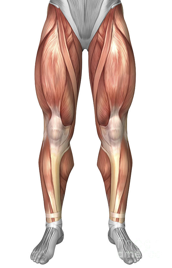
The bones of the leg are the femur, tibia, fibula and patella. The knee is a strong but flexible hinge joint. The accompanying muscle diagram reveals the position of the muscles of the lower legs in this pose. Observe the bones with a hand lens. Your leg bones are very large and strong to help support the weight of your body. This long bone connects with the knee at one end and the ankle at the other. The bones of your leg have roughened patches on their surfaces where muscles are attached. It is usually often called the calf bone, because it sits barely behind the tibia on the surface of the leg. Master leg and knee anatomy using our topic page. Vector isolated illustration of human arterial and venous circulatory system in leg anatomy.

The shoulder is made up of three bones:
Click now to learn more about the bones, muscles, and soft tissues of these regions at kenhub! Your leg bones are the longest and strongest bones in your body. The lower leg consists of two bones: Your leg bones are very large and strong to help support the weight of your body. Bones in the lower leg. The foot bones shown in this diagram are the talus, navicular, cuneiform, cuboid, metatarsals and calcaneus. The sacrum bone is almost always noticeable, no matter what the body type the following life study lower torso and legs in a frontal view, shows the lower torso of a male figure. Master leg and knee anatomy using our topic page. How to prepare baked chicken legs and thighs aka chicken leg quarters. The bones of your leg have roughened patches on their surfaces where muscles are attached. • the bones above the thigh are part of the hip and backbone of the chicken. The femur, or thigh bone, is the largest, heaviest, and strongest bone in the human body. Public domain image by jecowa at english wikipedia. At the distal end of the femur, two rounded condyles meet the tibia and fibula bones of the lower leg to form the knee joint. While their parts are similar in general, their structure has been adapted to differing functions.
It is also known as the calf bone, as it sits slightly behind the tibia on the outside of the leg. 409 x 400 jpeg 38 кб. • the bones above the thigh are part of the hip and backbone of the chicken. The second largest bone in body is the tibia, also called the shinbone. While their parts are similar in general, their structure has been adapted to differing functions.
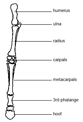
The humerus and the femur are corresponding bones of the arms and legs, respectively.
This long bone connects with the knee at one end and the ankle at the other. Leg muscle sport trauma and bone pain labeled diagram circulatory system anatomy. The shoulder is made up of three bones: Click now to learn more about the bones, muscles, and soft tissues of these regions at kenhub! At the distal end of the femur, two rounded condyles meet the tibia and fibula bones of the lower leg to form the knee joint. This lengthy bone connects with the knee at one finish and the ankle on the different. While their parts are similar in general, their structure has been adapted to differing functions. Time to jump right into the biggest and strongest bones in the human body. It is also known as the calf bone, as it sits slightly behind the tibia on the outside of the leg. Quizzes on human skeletal system anatomy, bone anatomy, and bone markings. A baby's skeleton typically consists of more individual bones.

Posting Komentar untuk "Bones In Leg Diagram"